
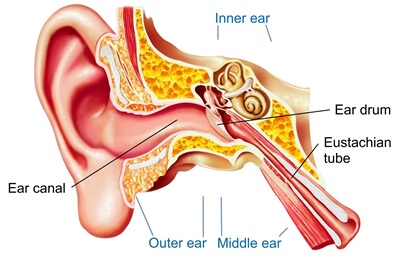
Types and Usage
The ear is the organ of hearing and, in mammals, balance. In mammals, the ear is usually described as having three parts—the outer ear, middle ear and the inner ear.
Outer ear:
The outer ear is the external portion of the ear and includes the fleshy visible pinna (also called the auricle), the ear canal, and the outer layer of the eardrum (also called the tympanic membrane).
The pinna consists of a single piece of elastic cartilage with a complicated relief on its inner surface and a fairly smooth configuration on its posterior surface. A tubercle, known as Darwin’s tubercle, is sometimes present, lying in the descending part of the helix and corresponding to the ear-tip of mammals. The earlobe consists of areola and adipose tissue. The symmetrical arrangement of the two ears allows for the localisation of sound. The brain accomplishes this by comparing arrival-times and intensities from each ear, in circuits located in the superior olivary complex and the trapezoid bodies which are connected via pathways to both ears.
Middle ear:
The middle ear lies between the outer ear and the inner ear. It consists of an air-filled cavity called the tympanic cavity and includes the three ossicles and their attaching ligaments; the auditory tube; and the round and oval windows. The ossicles are three small bones that function together to receive, amplify, and transmit the sound from the eardrum to the inner ear.
The three ossicles transmit sound from the outer ear to the inner ear. The malleus receives vibrations from sound pressure on the eardrum, where it is connected at its longest part (the manubrium or handle) by a ligament.
The round window allows for the fluid within the inner ear to move. As the stapes pushes the secondary tympanic membrane, fluid in the inner ear moves and pushes the membrane of the round window out by a corresponding amount into the middle ear. The ossicles help amplify sound waves by nearly 15–20 times.
Inner ear:
The inner ear sits within the temporal bone in a complex cavity called the bony labyrinth. A central area known as the vestibule contains two small fluid-filled recesses, the utricle and saccule.
The bony labyrinth refers to the bony compartment which contains the membranous labyrinth, contained within the temporal bone. The inner ear structurally begins at the oval window, which receives vibrations from the incus of the middle ear. Vibrations are transmitted into the inner ear into a fluid called endolymph, which fills the membranous labyrinth. The endolymph is situated in two vestibules, the utricle and saccule, and eventually transmits to the cochlea, a spiral-shaped structure. The cochlea consists of three fluid-filled spaces: the vestibular duct, the cochlear duct, and the tympanic duct. Hair cells responsible for transduction—changing mechanical changes into electrical stimuli are present in the organ of Corti in the cochlea.


