
Knowledge, Information and Experience
The ear has been adorned by earrings and other jewelry in numerous cultures for thousands of years, and has been subjected to surgical and cosmetic alterations.
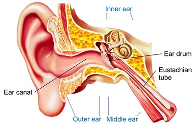
Structure:
The human ear consists of three parts—the outer ear, middle ear and inner ear. The ear canal of the outer ear is separated from the air-filled tympanic cavity of the middle ear by the eardrum. The middle ear contains the three small bones—the ossicles—involved in the transmission of sound, and is connected to the throat at the nasopharynx, via the pharyngeal opening of the Eustachian tube. The inner ear contains the otolith organs—the utricle and saccule—and the semicircular canals belonging to the vestibular system, as well as the cochlea of the auditory system.
Blood supply:
The blood supply of the ear differs according to each part of the ear.
The outer ear is supplied by a number of arteries. The posterior auricular artery provides the majority of the blood supply. The anterior auricular arteries provide some supply to the outer rim of the ear and scalp behind it.
The middle ear is supplied by the mastoid branch of either the occipital or posterior auricular arteries and the deep auricular artery, a branch of the maxillary artery.
The inner ear is supplied by the anterior tympanic branch of the maxillary artery; the stylomastoid branch of the posterior auricular artery; the petrosal branch of middle meningeal artery; and the labyrinthine artery, arising from either the anterior inferior cerebellar artery or the basilar artery.
Hearing:
Sound waves travel through the outer ear, are modulated by the middle ear, and are transmitted to the vestibulocochlear nerve in the inner ear. This nerve transmits information to the temporal lobe of the brain, where it is registered as sound.
Balance:
Providing balance, when moving or stationary, is also a central function of the ear. The ear facilitates two types of balance: static balance, which allows a person to feel the effects of gravity, and dynamic balance, which allows a person to sense acceleration.
Development:
During embryogenesis the ear develops as three distinct structures: the inner ear, the middle ear and the outer ear. Each structure originates from a different germ layer: the ectoderm, endoderm and mesenchyme.
After implantation, around the second to third week the developing embryo consists of three layers: endoderm, mesoderm and ectoderm. The first part of the ear to develop is the inner ear, which begins to form from the ectoderm around the 22nd day of the embryo’s development.
The ossicles (malleus, incus and stapes) normally appear during the first half of fetal development. The first two (malleus and incus) derive from the first pharyngeal arch and the stapes derives from the second.
Unlike structures of the inner and middle ear, which develop from pharyngeal pouches, the ear canal originates from the dorsal portion of the first pharyngeal cleft. It is fully expanded by the end of the 18th week of development.


