Tetralogy of Fallot (teh-TRAL-uh-jee of fuh-LOW) is a rare condition caused by a combination of four heart defects that are present at birth (congenital).
These defects, which affect the structure of the heart, cause oxygen-poor blood to flow out of the heart and to the rest of the body. Infants and children with tetralogy of Fallot usually have blue-tinged skin because their blood doesn’t carry enough oxygen.
Symptoms
Tetralogy of Fallot symptoms vary, depending on the amount of blood flow that’s blocked. Signs and symptoms may include:
- A bluish coloration of the skin caused by low blood oxygen levels (cyanosis)
- Shortness of breath and rapid breathing, especially during feeding or exercise
- Poor weight gain
- Tiring easily during play or exercise
- Irritability
- Prolonged crying
- Heart murmur
- Fainting
- An abnormal, rounded shape of the nail bed in the fingers and toes (clubbing)
Causes
Tetralogy of Fallot occurs as the baby’s heart is developing during pregnancy. Usually, the cause is unknown.
Tetralogy of Fallot includes four defects:
Narrowing of the lung valve (pulmonary valve stenosis). Narrowing of the valve that separates the lower right chamber of the heart (right ventricle) from the main blood vessel leading to the lungs (pulmonary artery) reduces blood flow to the lungs. The narrowing might also affect the muscle beneath the pulmonary valve. Sometimes, the pulmonary valve doesn’t form properly (pulmonary atresia).
A hole between the bottom heart chambers (ventricular septal defect). A ventricular septal defect is a hole in the wall (septum) that separates the two lower chambers of the heart (left and right ventricles). The hole causes oxygen-poor blood in the right ventricle to mix with oxygen-rich blood in the left ventricle. This causes inefficient blood flow and reduces the supply of oxygen-rich blood to the body. The defect eventually can weaken the heart.
Shifting of the body’s main artery (aorta). Normally the aorta branches off the left ventricle. In tetralogy of Fallot, the aorta is in the wrong position. It’s shifted to the right and lies directly above the hole in the heart wall (ventricular septal defect). As a result, the aorta receives a mix of oxygen-rich and oxygen-poor blood from both the right and left ventricles.
Thickening of the right lower heart chamber (right ventricular hypertrophy). When the heart’s pumping action is overworked, the muscular wall of the right ventricle becomes thick. Over time this might cause the heart to stiffen, become weak and eventually fail.
Risk Factors
While the exact cause of tetralogy of Fallot is unknown, some things might increase the risk of a baby being born with this condition. Risk factors for tetralogy of Fallot include:
- A viral illness during pregnancy, such as rubella (German measles)
- Drinking alcohol during pregnancy
- Poor nutrition during pregnancy
- A mother older than age 40
- A parent who has tetralogy of Fallot
- The presence of Down syndrome or DiGeorge syndrome in the baby
Complications
A possible complication of tetralogy of Fallot is infection of the inner lining of the heart or heart valve caused by a bacterial infection (infective endocarditis). Your or your child’s doctor may recommend taking antibiotics before certain dental procedures to prevent infections that might cause this infection.
People with untreated tetralogy of Fallot usually develop severe complications over time, which might result in death or disability by early adulthood.
Complications from tetralogy of Fallot surgery
While most babies and adults do well after open-heart surgery to repair tetralogy of
Fallot defects (intracardiac repair), long-term complications are common. Complications may include:
- Leaking pulmonary valve (chronic pulmonary regurgitation), in which blood leaks through the valve back into the pumping chamber (right ventricle)
- Leaking tricuspid valve
- Holes in the wall between the ventricles (ventricular septal defects) that may continue to leak after repair or may need re-repair
- Enlarged right ventricle or left ventricle that isn’t working properly
- Irregular heartbeats (arrhythmias)
- Coronary artery disease
- Enlargement of the ascending aorta (aortic root dilation)
- Sudden cardiac death
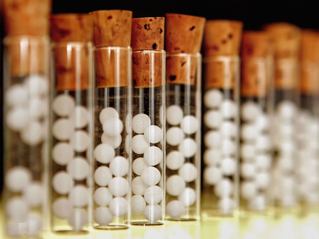
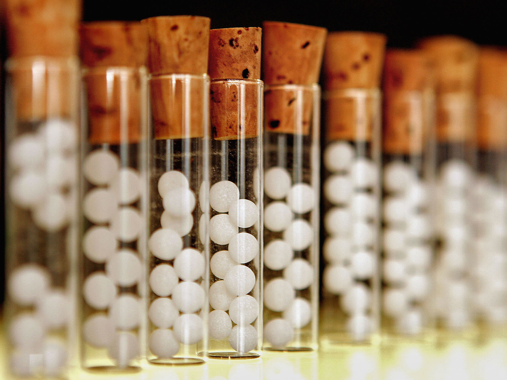
Homeopathic Treatment for Heart Hole
Homoeopathy is one of the most extensive holistic methods of medicine.
- Homoeopathy offers a significant complementary role in the treatment of several cardiovascular diseases.
- Homoeopathic remedies are effective in treating heart diseases, either as a therapeutic or as preventive medicine.
- The selection of treatment relies upon the principles of individualization and symptoms similarity by utilizing the holistic approach.
- It is the only way to regain the state of complete health by eliminating all the signs and symptoms the patient is suffering.
- For personalized and effective treatment, you should consult a highly qualified homoeopath in person.
There are various homoeopathic medicines used in treating Atrial Septal Defects, including:
- Phosphorus 30, 200
- Sulfur 200
- Calc Phos 6x
- Apis
- Lachesis


